

Anatomical Models
for Surgical Training
A reference compendium of high-fidelity, 3D-printed pathology simulators — engineered for the rehearsal, instruction, and study of foot and ankle surgery.
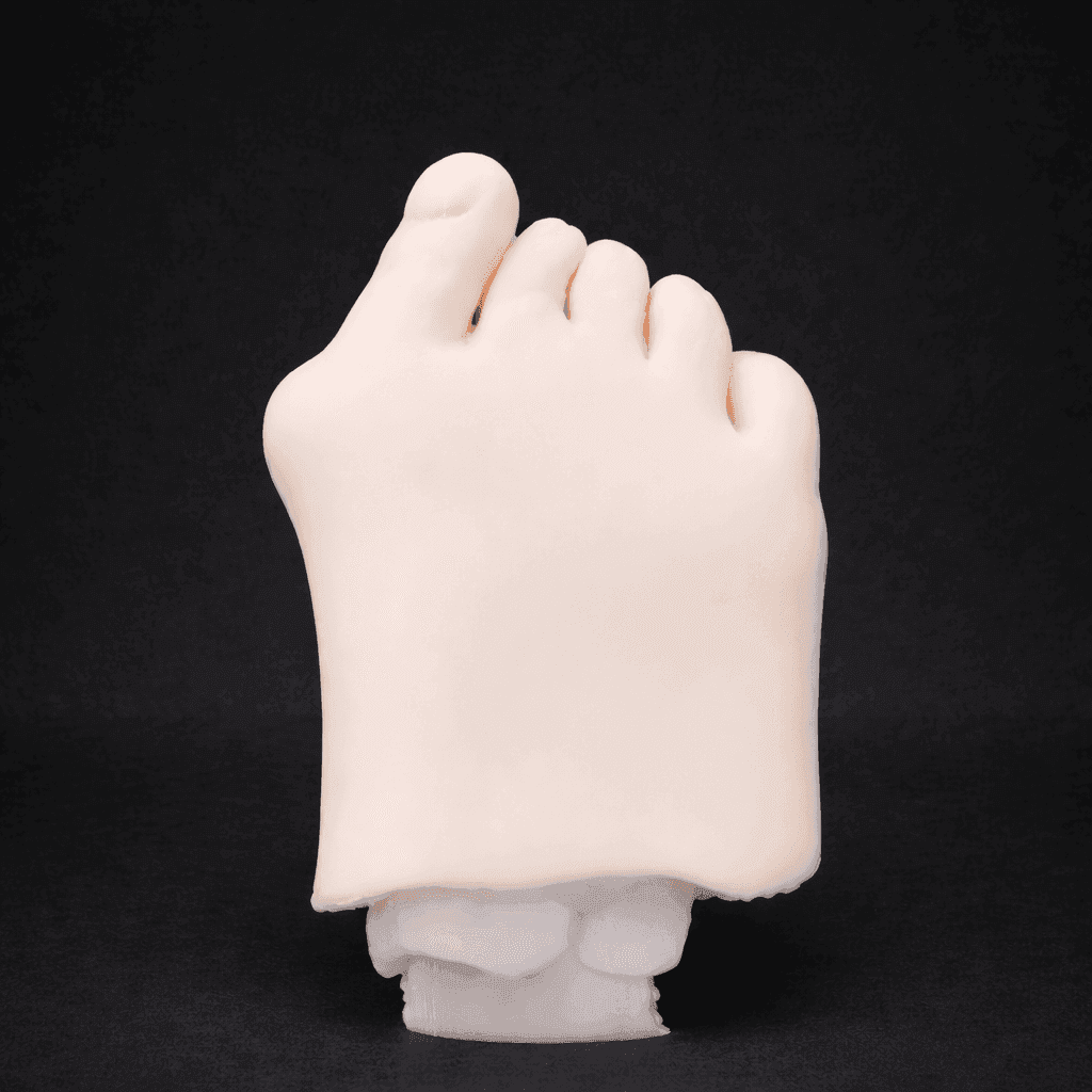
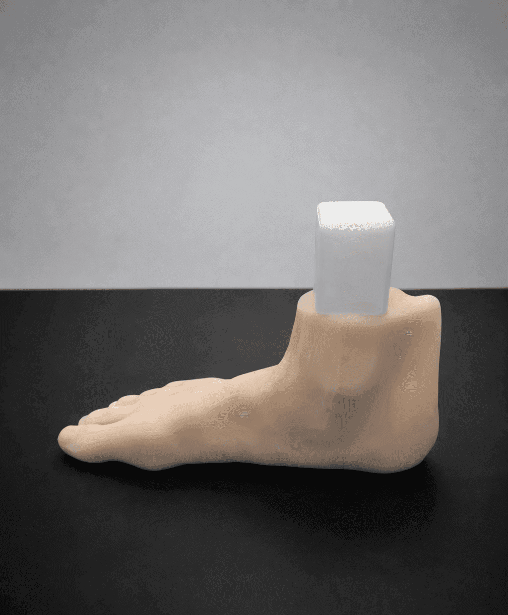
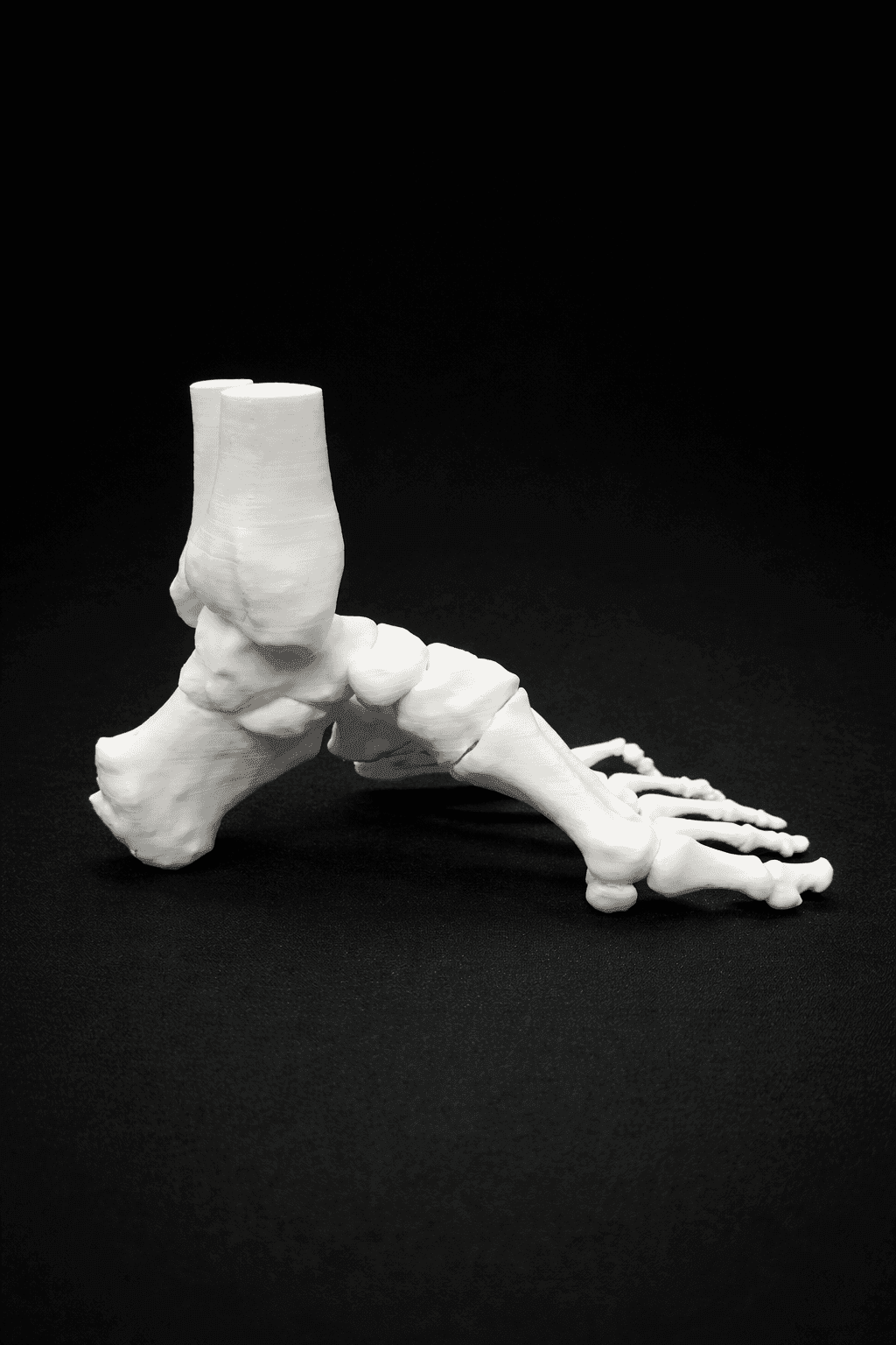
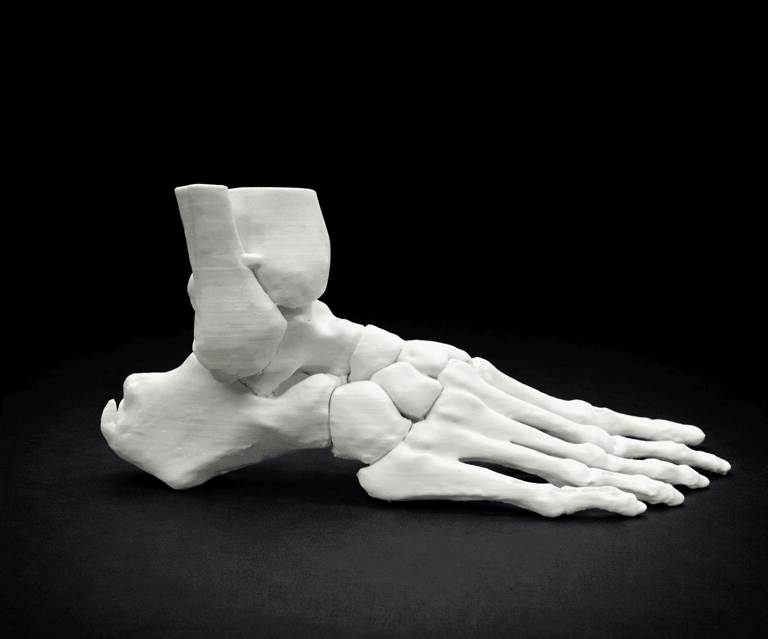
A reference instrument for pre-operative rehearsal, didactic instruction, and surgical study.
KIOPSI anatomical models are produced by additive manufacturing from medical-imaging data to faithfully reproduce the osseous and soft-tissue architecture of clinically encountered foot and ankle pathologies. Each reference is fabricated with material combinations selected to approximate the tactile and radiographic behavior of native tissues — supporting both open and image-guided rehearsal.
The catalog is intended for use by orthopaedic and podiatric surgeons, surgical residents, allied training programs, and medical device representatives in the context of pre-operative planning, technical instruction, instrument validation, and surgical skill development. Models are not intended for implantation.
Each reference in this volume corresponds to a discrete catalog number (REF) and is supplied with documentation of its material composition, dimensional envelope, and intended pathology.
- IHallux Valgus7 REF
- IIPes Planus4 REF
- IIIPes Cavus4 REF
- IVHaglund Deformity & Insertional Achilles Tendinopathy4 REF
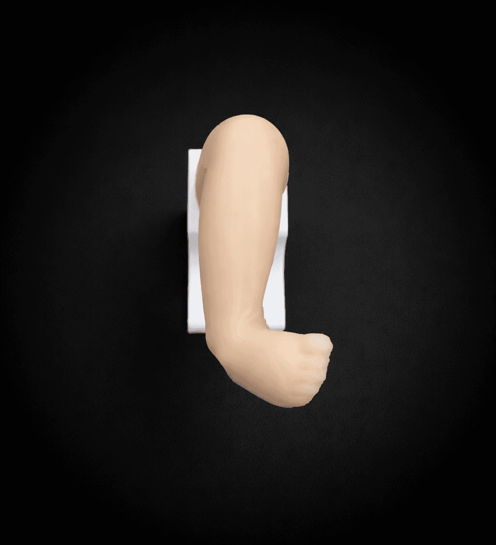
- VPes Equinovarus2 REF
- VIHallux Valgus & Pes Planus4 REF
- VIIHallux Valgus & Pes Planus & Haglund1 REF
- VIIITalus Osteochondritis Dissecans (OCD)1 REF
- IXOther1 REF
- AIndications for UseFront matter
- BProduct Catalog28 REF
- COrdering Information3 columns
- DTechnical Specifications & MaterialsReference
- EContact & InquiriesBack matter
- FColophonEnd
Hallux Valgus
Progressive lateral deviation of the great toe at the metatarsophalangeal joint, frequently accompanied by medial eminence formation and first-ray instability.
Opaque Skin – Radioopaque Bone – Mobile Joints
- Bone
- Radioopaque
- Skin
- Opaque
- Dimensions
- 20x11x6 cm
- Mass
- 350 g
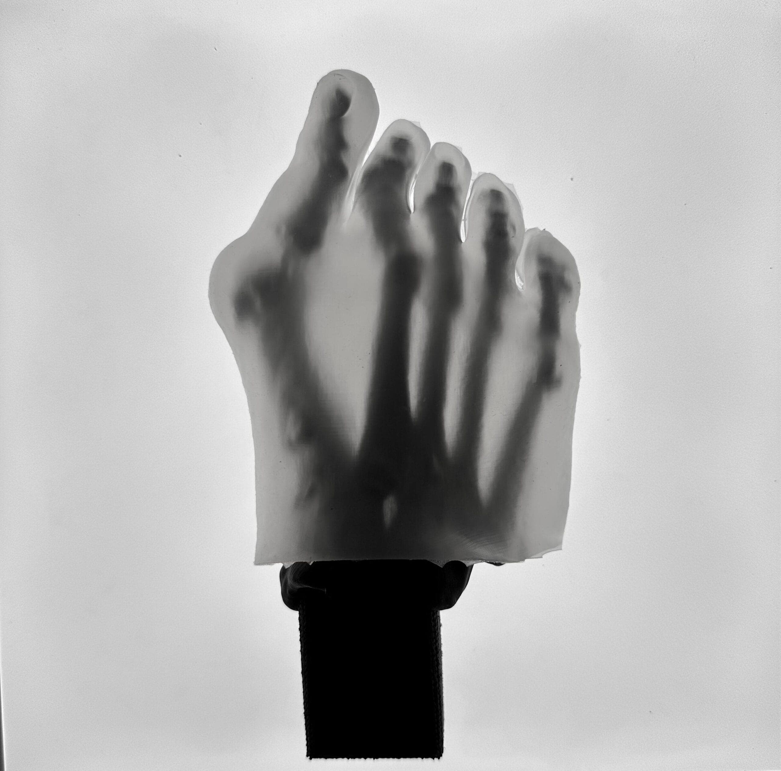
Transparent Skin – Radioopaque Black Bone – Mobile Joints
- Bone
- Radioopaque
- Skin
- Transparent
- Dimensions
- 20x11x6 cm
- Mass
- 350 g
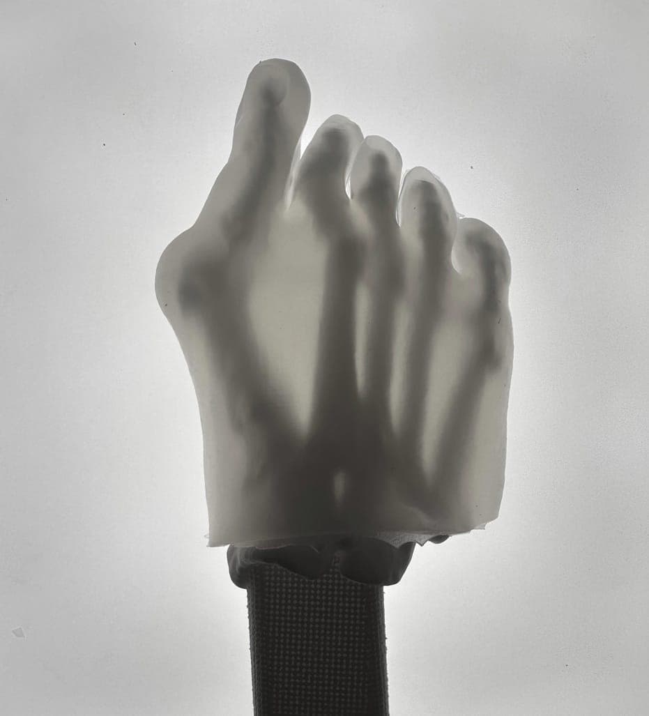
Transparent Skin – Radioopaque White Bone – Mobile Joints
- Bone
- Radioopaque
- Skin
- Transparent
- Dimensions
- 20x11x6 cm
- Mass
- 350 g
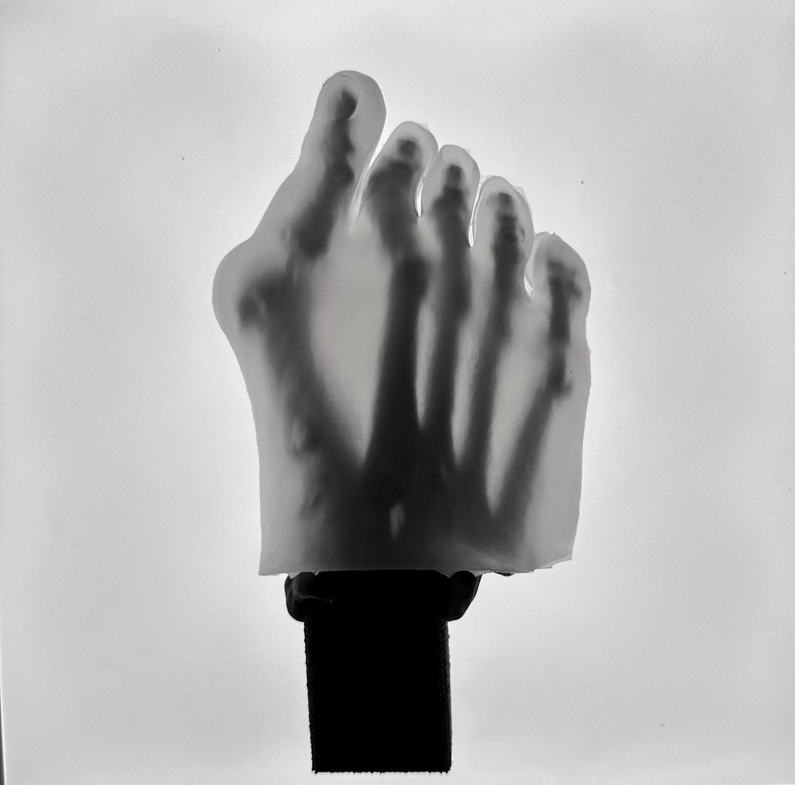
Transparent Skin – Radiolucent Black Bone – Mobile Joints
- Bone
- Radiolucent
- Skin
- Transparent
- Dimensions
- 20x11x6 cm
- Mass
- 350 g
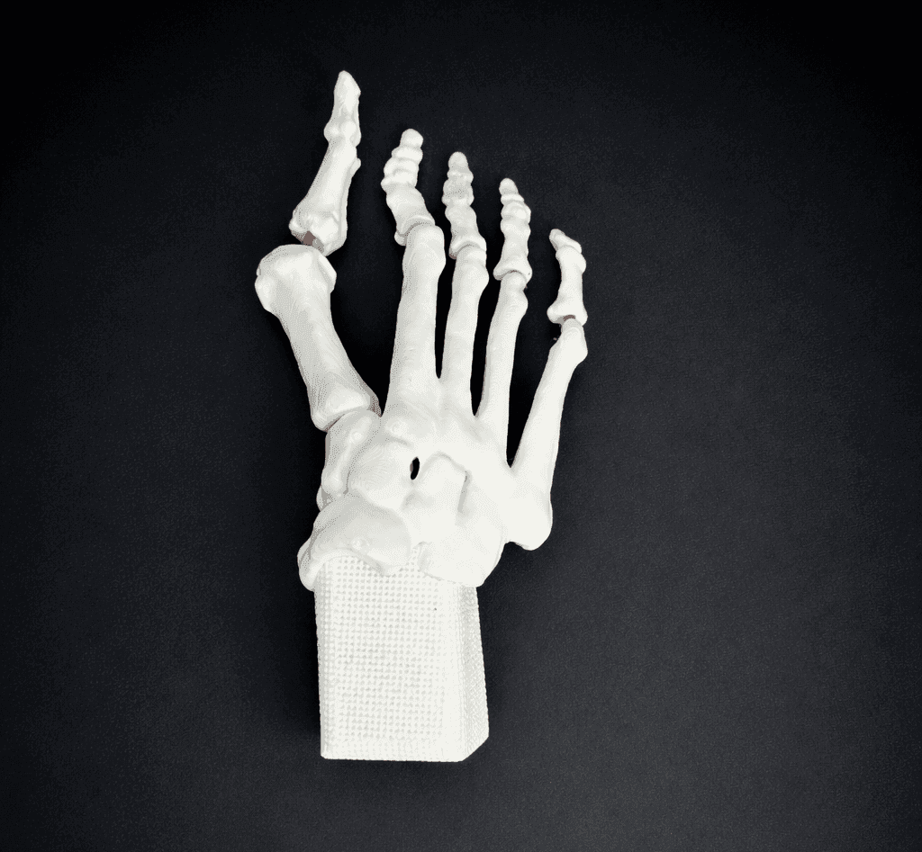
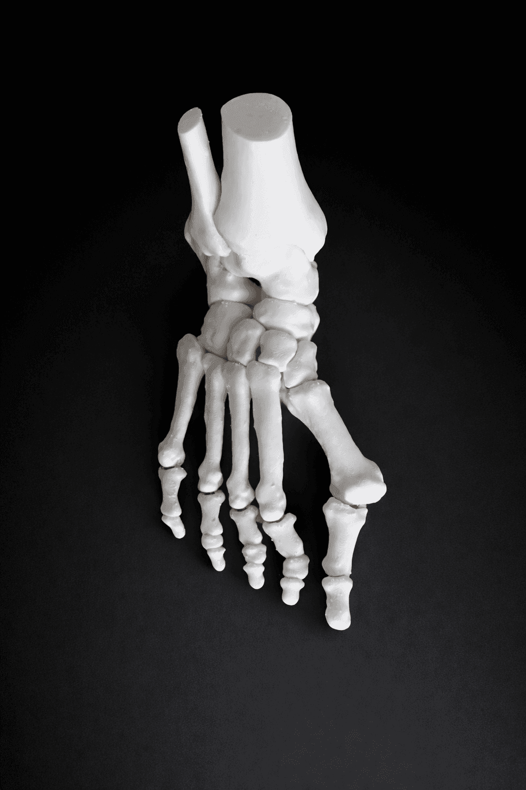
Bone Model – Mobile Joints
- Bone
- -
- Skin
- -
- Dimensions
- 19x19x4 cm
- Mass
- 120 g
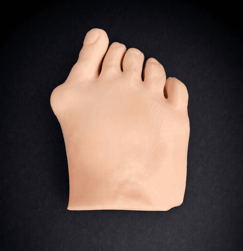
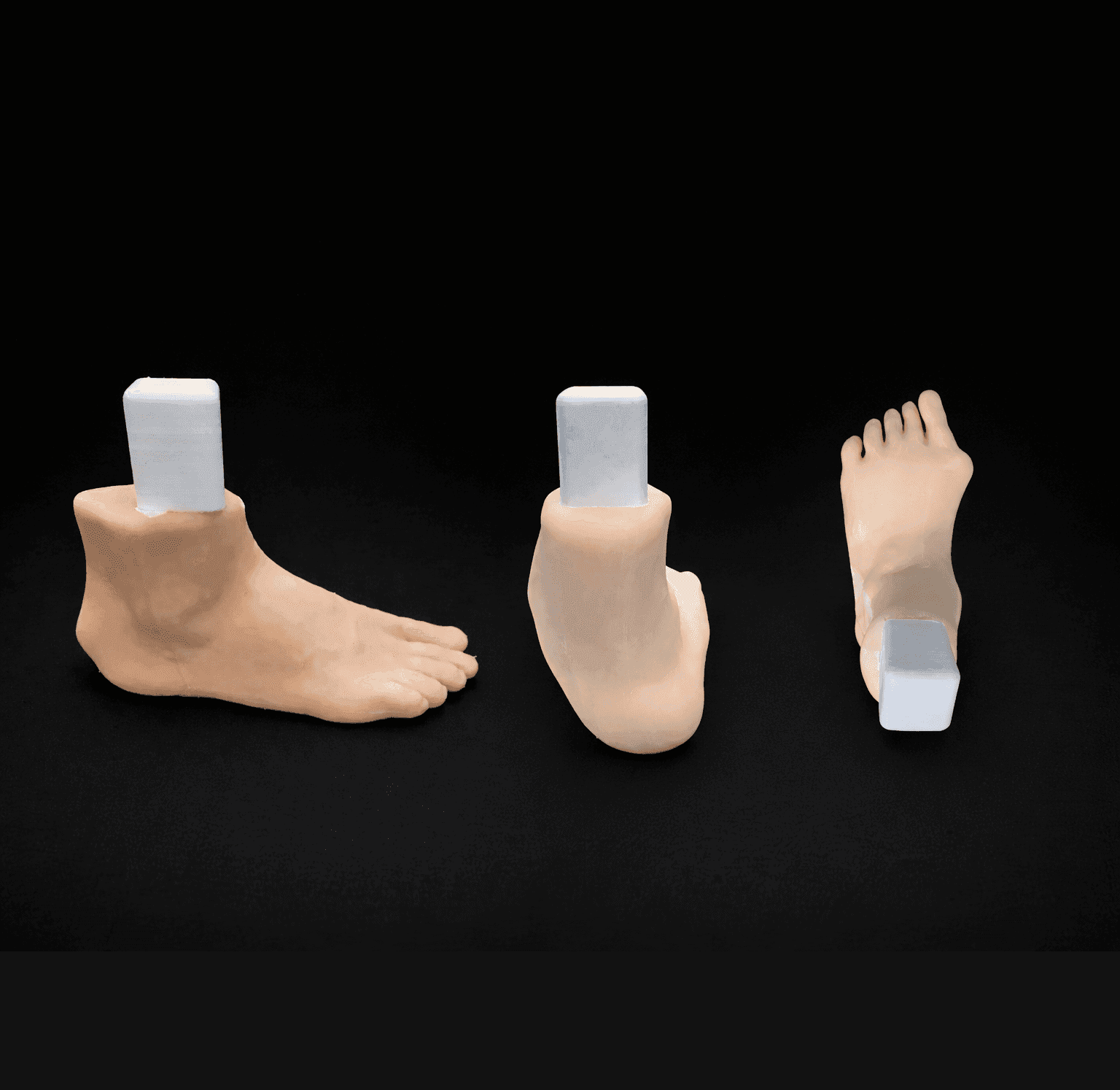
Modular Skin Part Compatible with Hallux Valgus Bone Model (Opaque)
- Bone
- -
- Skin
- Opaque
- Dimensions
- 14x11x5 cm
- Mass
- 260 g
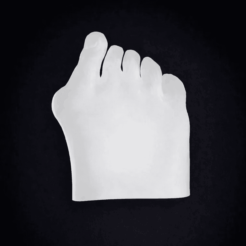
Modular Skin Part Compatible with Hallux Valgus Bone Model (Transparent)
- Bone
- -
- Skin
- Transparent
- Dimensions
- 14x11x5 cm
- Mass
- 260 g
Pes Planus
Loss of the medial longitudinal arch producing a flatfoot deformity, classified as flexible or rigid depending on hindfoot mobility.
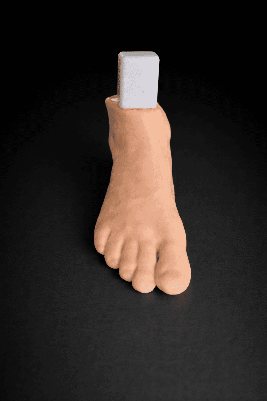
Radioopaque Bone – Opaque Skin – Mobile Joints (including Evans & Cotton wedges)
- Bone
- Radioopaque
- Skin
- Opaque
- Dimensions
- 28x17x11 cm
- Mass
- 860 g
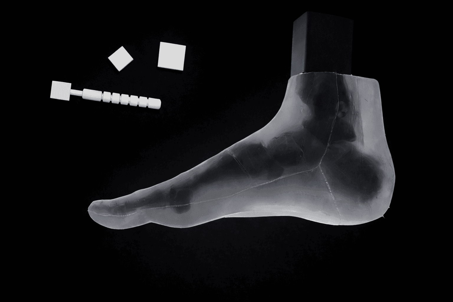
Transparent Skin – Radiolucent Black Bone – Mobile Joints (including Evans & Cotton wedges)
- Bone
- Radiolucent
- Skin
- Transparent
- Dimensions
- 28x17x11 cm
- Mass
- 860 g
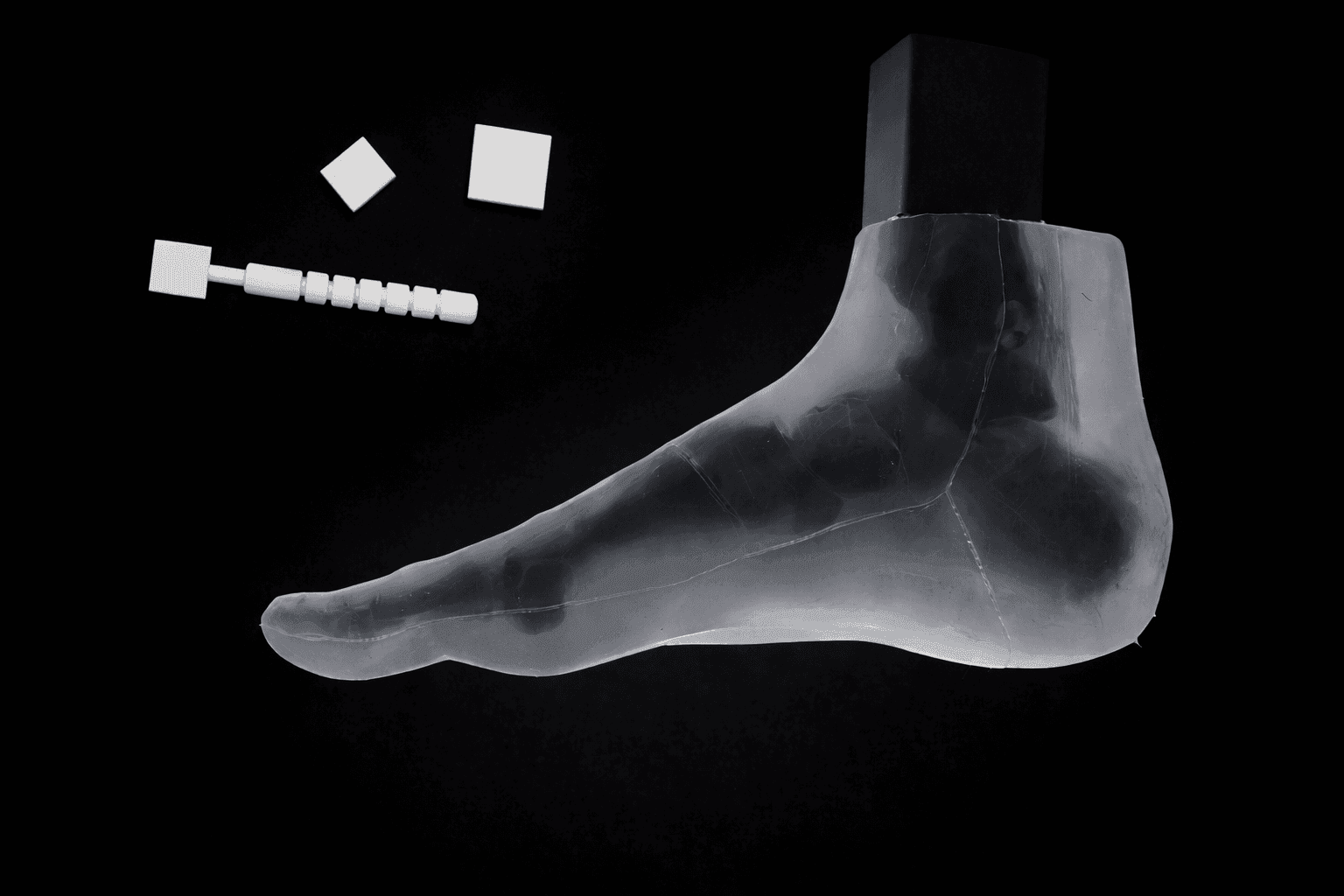
Transparent Skin – Radioopaque Black Bone – Mobile Joints (including Evans & Cotton wedges)
- Bone
- Radioopaque
- Skin
- Transparent
- Dimensions
- 28x17x11 cm
- Mass
- 860 g
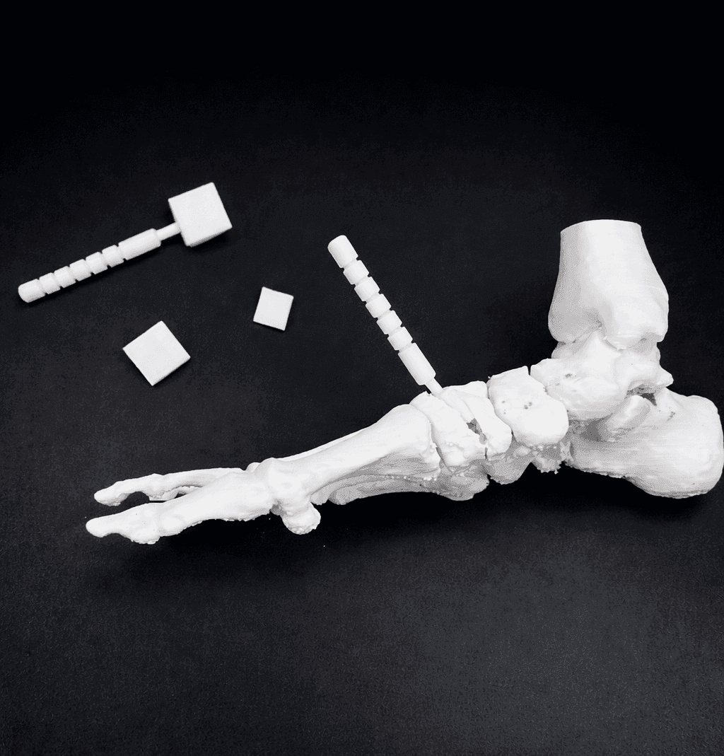
Bone Model – Mobile Tarsal Joints (including Evans & Cotton wedges)
- Bone
- Radioopaque
- Skin
- -
- Dimensions
- 22x13x8 cm
- Mass
- 140 g
Pes Cavus
Abnormally high medial longitudinal arch, commonly associated with hindfoot varus and forefoot equinus deformities.
Bone Model
- Bone
- -
- Skin
- -
- Dimensions
- 22x13x8 cm
- Mass
- 140 g
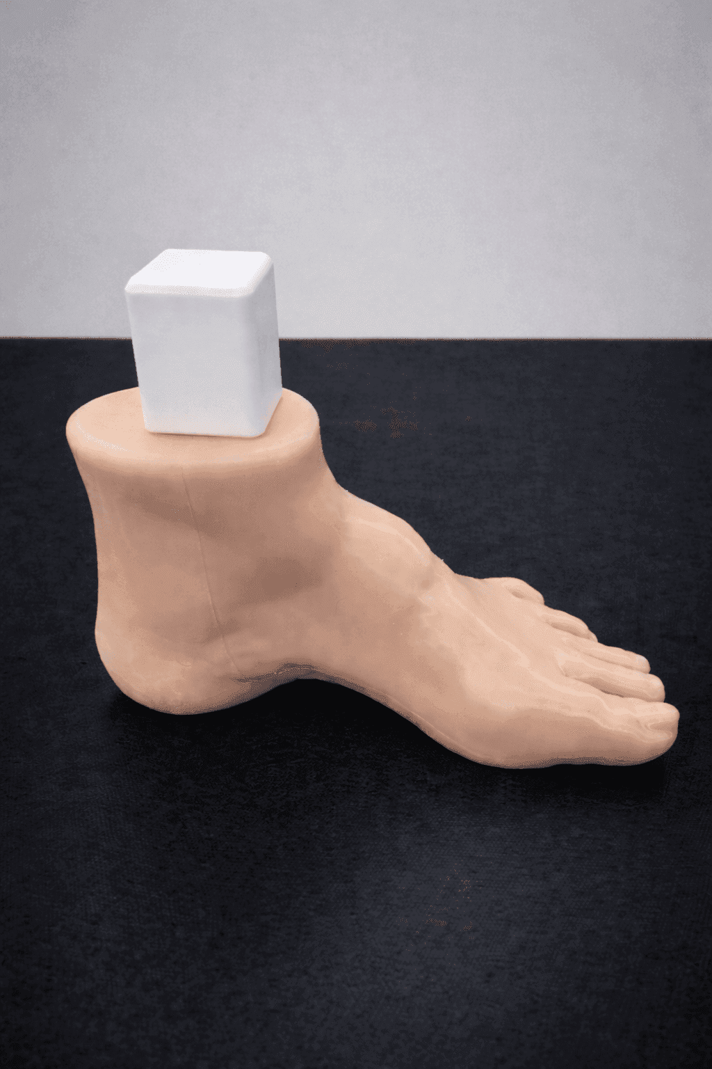
Radioopaque Bone – Opaque Skin
- Bone
- Radioopaque
- Skin
- Opaque
- Dimensions
- 23x18x10 cm
- Mass
- 970 g
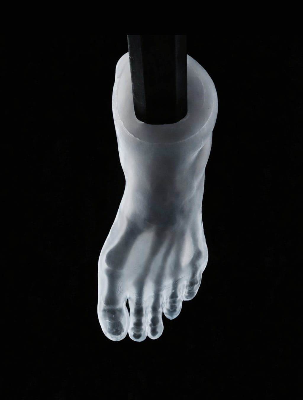
Transparent Skin – Radiolucent Black Bone
- Bone
- Radiolucent
- Skin
- Transparent
- Dimensions
- 23x18x10 cm
- Mass
- 970 g
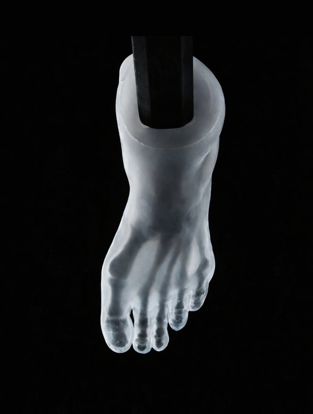
Transparent Skin – Radioopaque Black Bone
- Bone
- Radioopaque
- Skin
- Transparent
- Dimensions
- 23x18x10 cm
- Mass
- 970 g
Haglund Deformity & Insertional Achilles Tendinopathy
Anatomically accurate representation of the referenced pathology, reproduced to support pre-operative planning, surgical rehearsal, and resident education.
Bone Model
- Bone
- -
- Skin
- -
- Dimensions
- 22x13x8 cm
- Mass
- 140 g
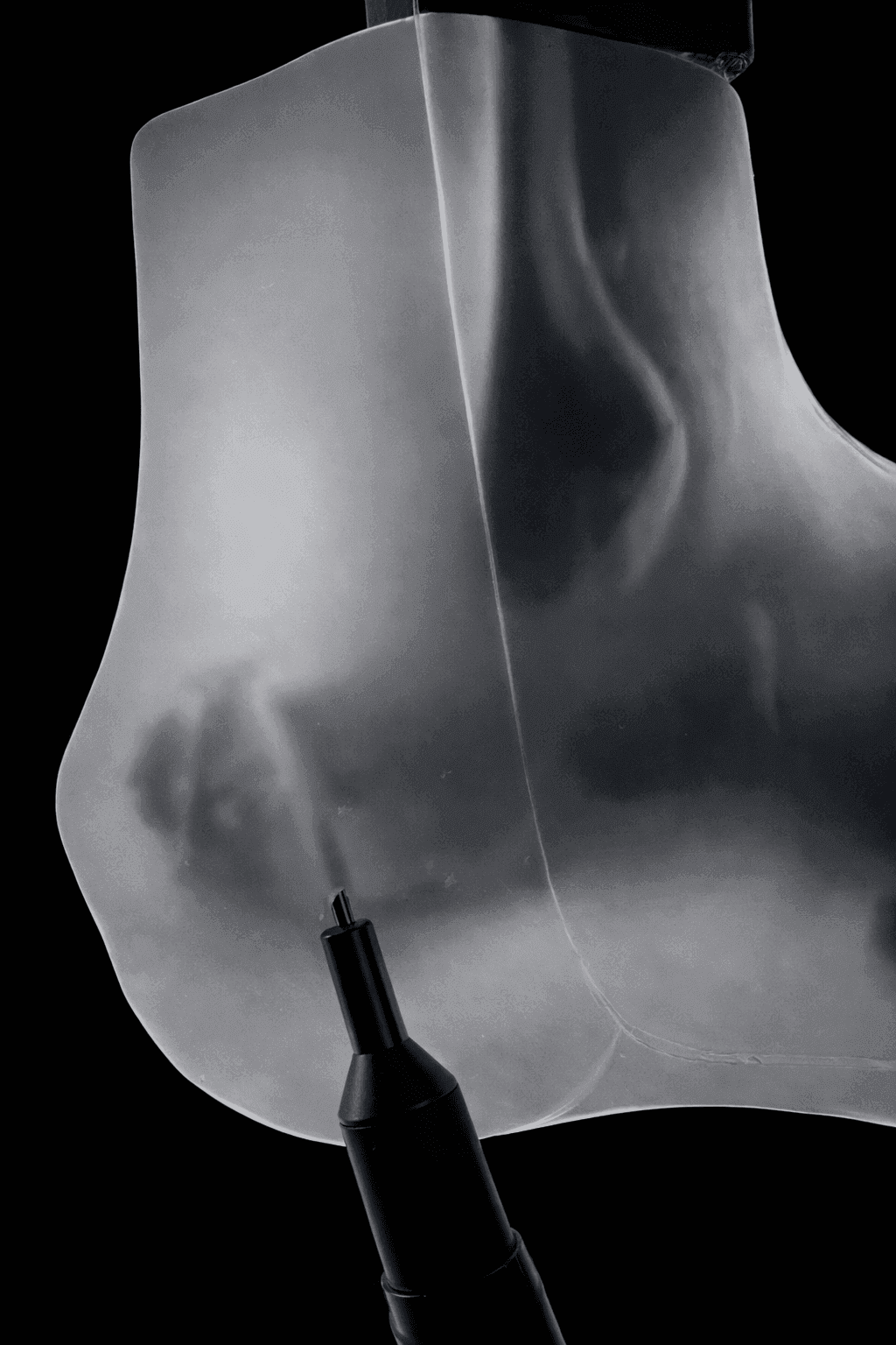
Transparent Skin – Radioopaque Black Bone
- Bone
- Radioopaque
- Skin
- Transparent
- Dimensions
- 23x17x10 cm
- Mass
- 810 g
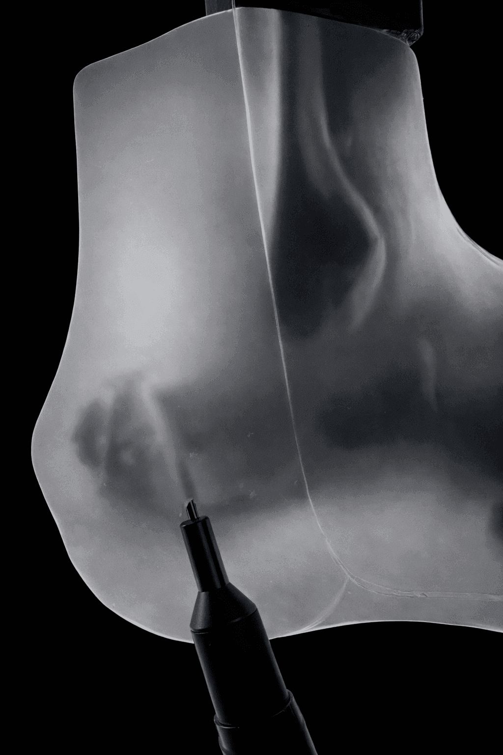
Transparent Skin – Radiolucent Black Bone
- Bone
- Radiolucent
- Skin
- Transparent
- Dimensions
- 23x17x10 cm
- Mass
- 810 g
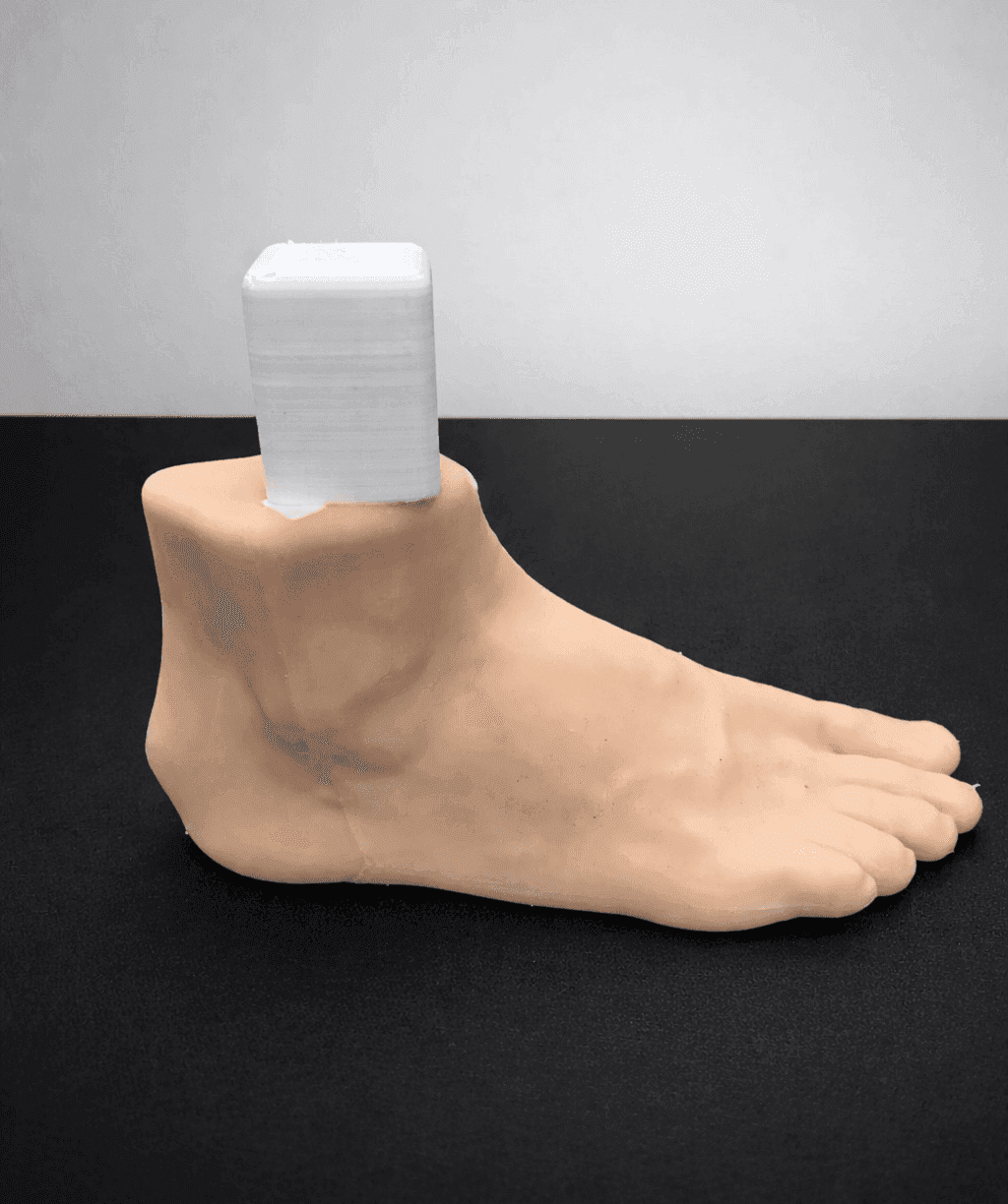
Radioopaque Bone – Opaque Skin
- Bone
- Radioopaque
- Skin
- Opaque
- Dimensions
- 23x17x10 cm
- Mass
- 810 g
Pes Equinovarus
Anatomically accurate representation of the referenced pathology, reproduced to support pre-operative planning, surgical rehearsal, and resident education.
Club Foot (Pes Equinovarus) – Uncorrected
- Bone
- Radiolucent
- Skin
- Opaque
- Dimensions
- 15x15x5 cm
- Mass
- 400 g
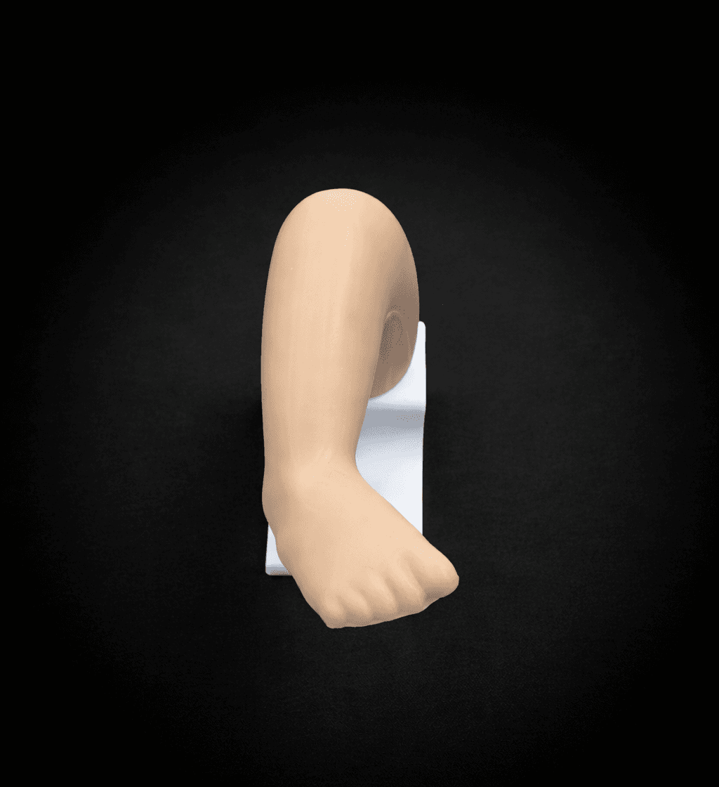
Club Foot (Pes Equinovarus) – Partially Corrected
- Bone
- Radiolucent
- Skin
- -
- Dimensions
- 15x15x5 cm
- Mass
- 400 g
Hallux Valgus & Pes Planus
Anatomically accurate representation of the referenced pathology, reproduced to support pre-operative planning, surgical rehearsal, and resident education.
2 Pathology in 1 Foot – Hallux Valgus & Pes Planus – Bone Model – Mobile Joints
- Bone
- -
- Skin
- -
- Dimensions
- 22x11x10 cm
- Mass
- 160 g
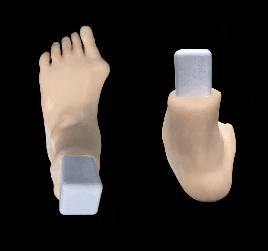
2 Pathology in 1 Foot – Hallux Valgus & Pes Planus – Radioopaque Bone – Opaque Skin – Mobile Joints
- Bone
- Radioopaque
- Skin
- Opaque
- Dimensions
- 23x15x11 cm
- Mass
- 980 g
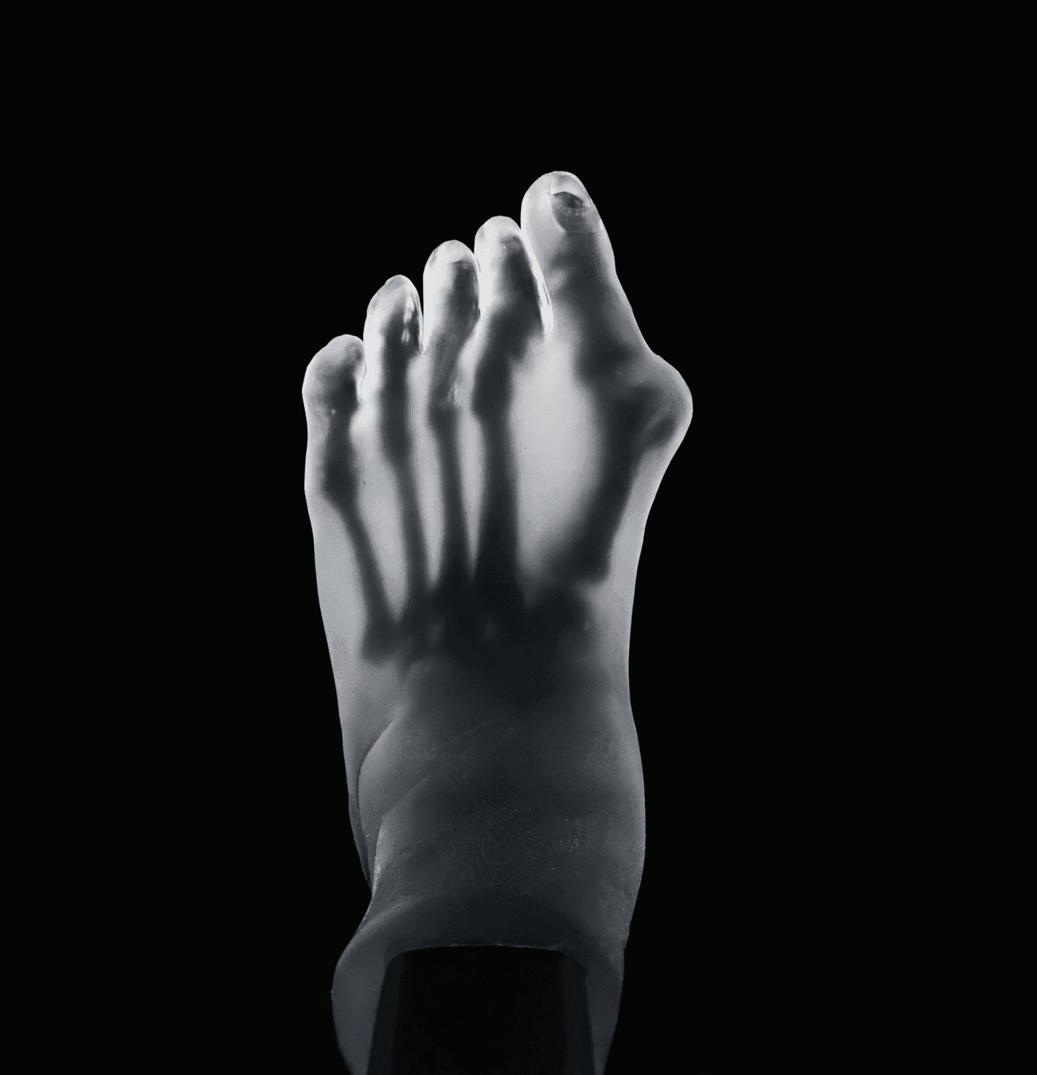
2 Pathology in 1 Foot – Hallux Valgus & Pes Planus – Transparent Skin – Radiolucent Bone – Mobile Joints
- Bone
- Radiolucent
- Skin
- Transparent
- Dimensions
- 23x15x11 cm
- Mass
- 980 g
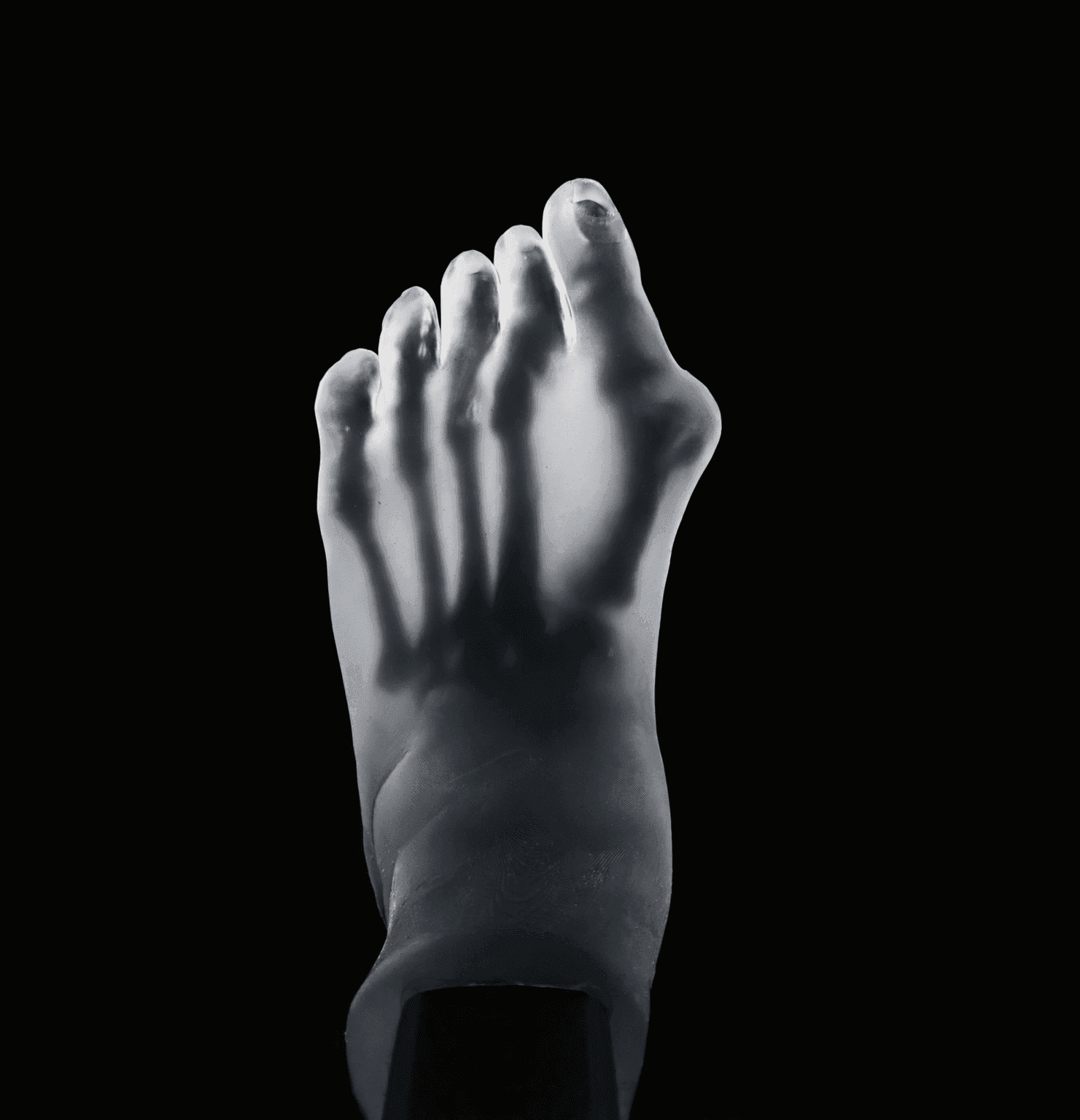
2 Pathology in 1 Foot – Hallux Valgus & Pes Planus – Transparent Skin – Radioopaque Bone – Mobile Joints
- Bone
- Radioopaque
- Skin
- Transparent
- Dimensions
- 23x15x11 cm
- Mass
- 980 g
Hallux Valgus & Pes Planus & Haglund
Anatomically accurate representation of the referenced pathology, reproduced to support pre-operative planning, surgical rehearsal, and resident education.
3 Pathology in 1 Foot – Hallux Valgus & Pes Planus & Haglund – Radioopaque Bone – Opaque Skin – Mobile Joints
- Bone
- Radioopaque
- Skin
- Opaque
- Dimensions
- 23x15x11 cm
- Mass
- 980 g
Talus Osteochondritis Dissecans (OCD)
Anatomically accurate representation of the referenced pathology, reproduced to support pre-operative planning, surgical rehearsal, and resident education.
Talus OCD – Ankle Arthroscopy Trainer
- Bone
- -
- Skin
- -
- Dimensions
- 29x19x12 cm
- Mass
- 1000 g
Other
Anatomically accurate representation of the referenced pathology, reproduced to support pre-operative planning, surgical rehearsal, and resident education.
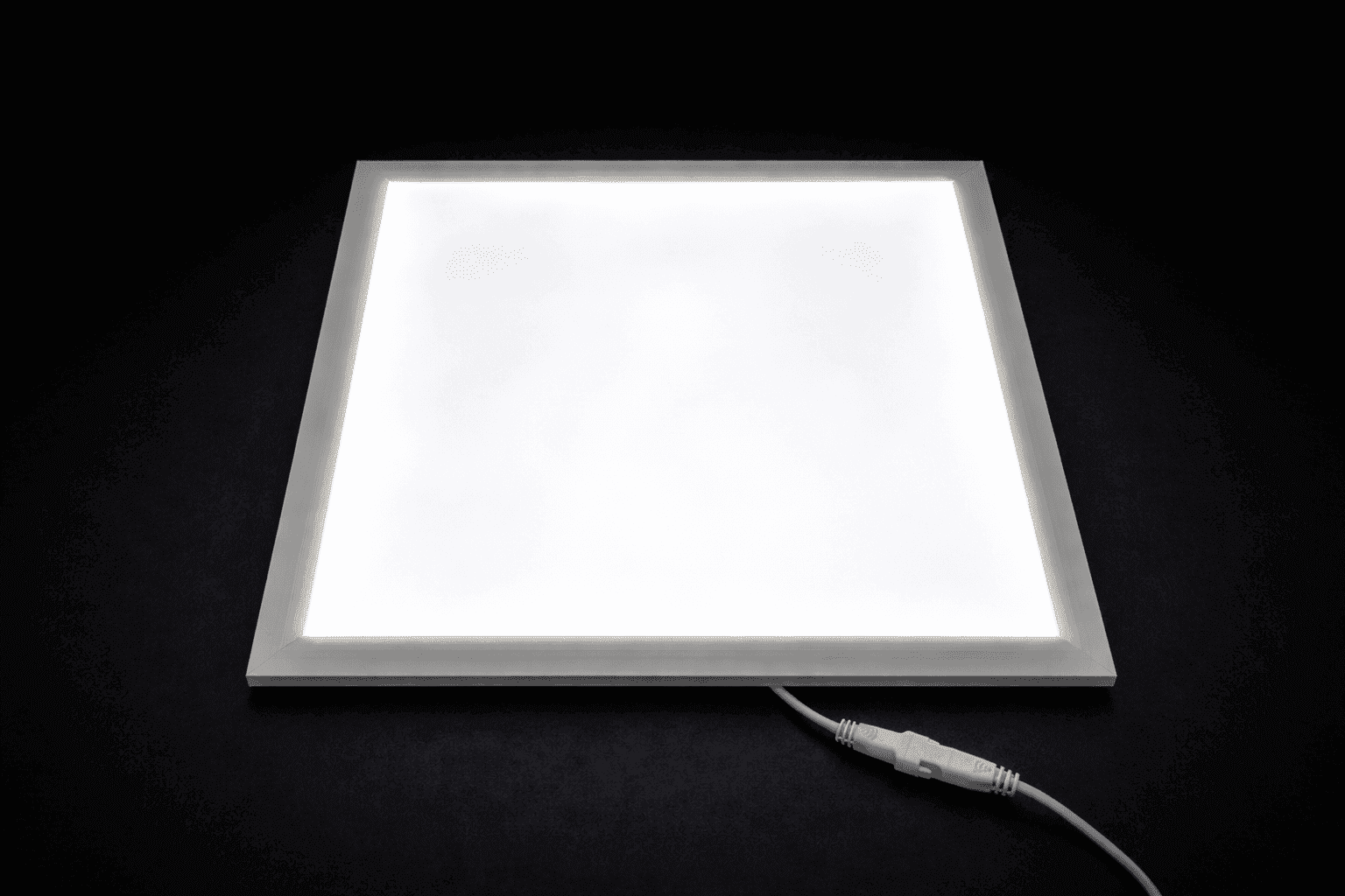
KIO-Ray – Advanced Visualization Platform for Transparent Models
- Bone
- -
- Skin
- -
- Dimensions
- 30x30x3 cm
- Mass
- 270 g
- Reference each model by its REF / Order Code as listed.
- Submit purchase orders via email to info@kiopsi.com.
- Indicate desired bone and skin variant where applicable.
- Institutional and academic purchase orders are accepted.
- Quotations are valid for thirty (30) days from issue.
- Standard catalog models: 10 — 14 business days.
- Variant configurations: 14 — 21 business days.
- Custom imaging-derived models: by quotation.
- Expedited fabrication available upon request.
- Shipping worldwide; duties and taxes by destination.
- Tiered pricing available for orders of five units or greater.
- Educational programs eligible for institutional discount.
- Distributor inquiries welcomed in unrepresented territories.
- Industry partnerships for instrument validation studies.
- Contact info@kiopsi.com for terms.
Osseous structures are fabricated from photopolymer resin systems formulated for either radiographic opacity or radiolucency. Soft-tissue envelopes use elastomeric resins tuned to approximate dermal compliance.
| Component | Variant | Behavior |
|---|---|---|
| Bone | Radioopaque (RO) | Visible under fluoroscopy |
| Bone | Radiolucent (RL) | Permits open instrumentation |
| Skin | Opaque (O) | Surface anatomy fidelity |
| Skin | Transparent (T) | Permits visual tracking |
Radioopaque references are suitable for use under C-arm fluoroscopy, mobile radiography, and computed tomography for the purposes of rehearsal and image-guided technique instruction. Radiolucent references are recommended where the surgeon requires unimpeded radiographic visualization of instrumentation.
Models are intended for single educational or rehearsal use unless otherwise indicated. Store at ambient room temperature, away from prolonged exposure to direct sunlight. Clean external surfaces with isopropyl alcohol if required. Models are not intended for autoclaving or for any form of in-vivo or implantation use.
KIOPSI anatomical models are classified under HTS 9023 as instructional models not designed for any other purpose. They are intended exclusively for surgical education, training, and pre-operative rehearsal. Models are not regulated medical devices and are not intended for diagnostic or therapeutic application.
Volume I · Edition I · 2026
Issued 2026-10-12